A rapid image cytometric analysis method for phagocytosis using Celigo Imaging Cytometer
Leo L. Chan, Scott Cribbes, Sarah Kessel, Olivier Dery, Dmitry Kuksin, and Jean Qiu
Phagocytosis is an essential process of the immune system to eliminate cellular debris and pathogens. It is a specific form of endocytosis involving vesicular internalization. Phagocytic cells such as macrophages can be
attracted to pathogens or cellular debris and engulf the material to be trapped in an internal vesicle called phagosome. The phagosome would fuse with the lysosome to form phagolysosome, where enzymes and toxic
peroxides can digest the pathogen or cellular debris at a low pH value. Traditionally, phagocytosis can be qualitatively examined via standard optical microscopy to observe the engulfment of bacteria or cellular debris. In
addition, phagocytosis can also be quantitatively measured by incubating phagocytes with particles bound with serum or IgG. After incubation, the excess particles are washed away, leaving only phagocytes with engulfed
particles. These cells are lysed to release the engulfed material, which can be examined using microplate reader or western blot can also be utilized to quantify the level of phagocytosis. However, these methods are timeconsuming and requires experienced researchers to obtain meaningful results. By using the Celigo Imaging Cytometer, phagocytosis can be measured using a pH sensitive label called pHrodo. The labels will only fluoresce when they are trapped in the phagolysosome where the pH value low. Therefore, by measuring the amount of fluorescence or percentage of cells fluorescing, one can use the Celigo to determine the fluorescent
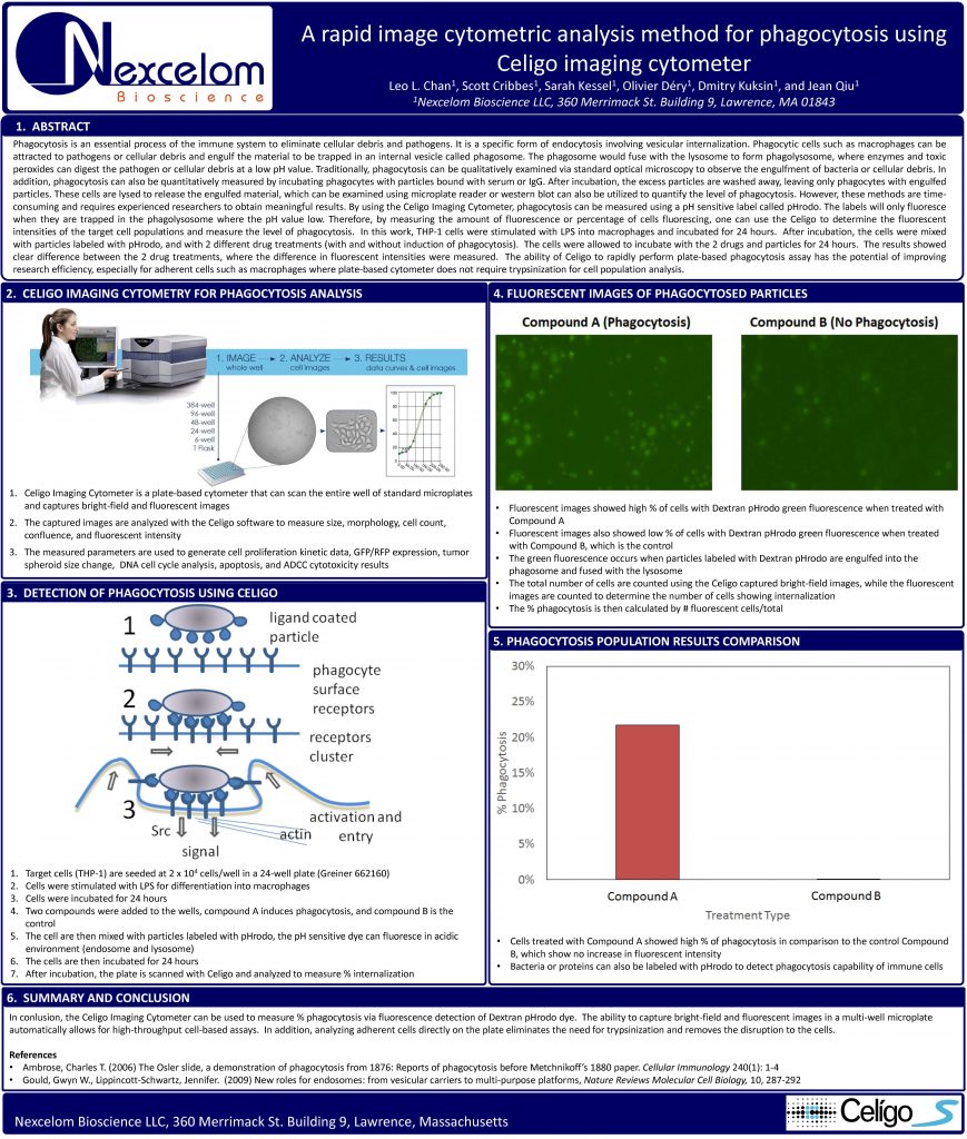
intensities of the target cell populations and measure the level of phagocytosis. In this work, THP-1 cells were stimulated with LPS into macrophages and incubated for 24 hours. After incubation, the cells were mixed with particles labeled with pHrodo, and with 2 different drug treatments (with and without induction of phagocytosis). The cells were allowed to incubate with the 2 drugs and particles for 24 hours. The results showed clear difference between the 2 drug treatments, where the difference in fluorescent intensities were measured. The ability of Celigo to rapidly perform plate-based phagocytosis assay has the potential of improving
research efficiency, especially for adherent cells such as macrophages where plate-based cytometer does not require trypsinization for cell population analysis.