Bright field and Fluorescent Image Analysis for Screening Applications using Celigo
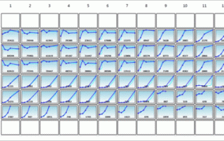
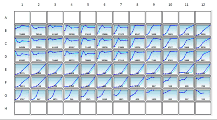
Generate growth curves over time and monitor cell counts and confluence at the individual well level.
Generate growth curves over time and monitor cell counts and confluence at the individual well level.
It's White Paper Wednesday! Read our featured white paper: Detection of Multiplexed GFP Reporters in Primary Articular Chondrocyte Cultures Using Cellometer Vision Image Cytometer In this work, we have developed an image cytometry method for detecting and monitoring the cell expansion and differentiation of articular chondrocytes in primary culture. First, the feasibility of utilizing image cytometry for detection of fluorescent is shown by comparing measured fluorescent positive cell populations to flow cytometry. Next, articular chondrocyte cultures were established in multi-well plates from either single or Cyan/eGFP double reporter mouse lines and grown for 20 days to test the utility of the [...]
It's White Paper Wednesday! Read our featured white paper:The Historical Development of the Hemacytometer The hemacytometer has been an essential tool for hematologists, medical practitioners, and biologists for over a century. Depending on where it is being used, the word has multiple spellings such as hemacytometer, hemocytometer, haemacytometer, or haemocytometer, but for consistency purposes the word “hemacytometer” will be used in this review. The prefix “hema”, “hemo”, “haema”, or “haemo” means blood, while “cytometer” meant a device to measure cells. The device was initially used by medical practitioners to analyze patient blood samples, which was the initial spark that created [...]
Introduction The oncogene c-Myc is frequently overexpressed in ovarian carcinomas [1]. The small molecule c-Myc inhibitor, 10058-F4, was investigated here to evaluate its effects on ovarian cancer cell growth as well as its mechanism of action [2]. The inhibitor’s impact on apoptosis, cell cycle, cell colony formation, ROS generation, and cell viability were examined in Hey and SKOV3 cell lines. These findings suggest that small molecule c-Myc inhibitors may be a promising strategy for future ovarian cancer therapies. Materials and Methods Cell cycle analysis Hey and SKOV3 cell lines (both human ovarian cancer) were plated in 96-well plates and treated [...]
Introduction A diverse family of cytokines, Type I interferons (IFNs), is a group of proteins responsible for antiviral, antiproliferative, and immunomodulatory functions [1, 2]. Treating cells with Type I IFNs induces autophagy, a cellular recycling process that is considered a key cell survival strategy [3]. It is well known that Type I IFNs work through the JAK-STAT pathway, but recent evidence suggests that the MAP kinase pathway can affect the expression of IFN-regulated genes [4, 5]. Researchers demonstrated that IFN-induced autophagy affects cell cycle as well as cellular proliferation in a variety of cell lines. Because autophagy plays a major [...]
Introduction Preclinical models to evaluate therapeutic efficacy in the area of oncology have proven to be a challenge. The standard of preclinical assessment as always been immunosuppressed mice, but that model has not reliably predicted clinical efficacy [1]. Immunocompetent genetically engineered mice with tumor allograft models seem more promising, and yet reporter tags such as GFP and luciferase are targeted by competent immune systems, which may interfere with tumor growth and the response to therapeutics. Here, a collaborative group of researchers has created a reporter-tolerized genetically engineered mouse model, dubbed “Glowing Head” (GH), by targeting a luciferase-GFP reporter into the [...]
Introduction The B-cell lymphoma-2 (BCL-2) family of proteins includes many key regulators of cell survival, cell death, and apoptosis. The deregulation of certain BCL-2 proteins is one of the first steps towards tumorigenesis and subsequent therapeutic resistance [1-4]. Targeting these BCL-2 family members is therefore an important area of oncology research [2, 4, 5]. Here, a collaboration of researchers has explored the targeting of these BCL-2 proteins with a BCL-2 binding peptide known as NuBCP-9 (FSRSLHSLL), which induces a conformational change in BCL-2 that alters the protein’s function from cell protection to cell killing. Researchers paired NuBCP-9 with iron oxide-based [...]
The entire family of tumor protein p53 (TP53) enhances functions such as apoptosis and autophagy in normal cellular functioning. TP53 is a tumor repressor gene that is often inactivated in human cancers. Reactivating p53 has proven difficult to achieve therapeutically, however. Researchers at MD Anderson Cancer Center are investigating other members of the p53 pathway in order to elucidate new therapeutic options to suppress p53-deficient tumor growth. ΔN isoforms of two members of the p53 family, p63 and p73, are usually overexpressed in cancers and these isoforms (which lack the acidic transactivation domain) act on p53 in a dominant-negative fashion, [...]
Nexcelom hosts free, live monthly training webinars for our Cellometer instruments. Our webinars cover a variety of instruments, cell types and applications. We have taken our recorded webinars and added them to our YouTube channel! We've created a playlist entitled "Cellometer Training Webinars" and the list will continue to grow as we release more. This is a great resource for you and new lab team members to get additional training on your Cellometer instrument. We host new training webinars each month, so revisit our YouTube channel from time to time to see new content as it's produced and added.
Introduction to Nanoparticle-Based Therapy for Triple-Negative Breast Cancer Tia Harmon and Dr. Ruben Gonzalez-Perez from the Morehouse School of Medicine and Emory University utilized the imaging and analysis capabilities of the Cellometer Vision to investigate leptin peptide receptor antagonist-conjugated nanoparticles in order to inhibit leptin signaling, a key pathway that promotes growth and survival of triple-negative breast cancer (TNBC) cells. It was hypothesized that these nanoparticles would first stop the expression of leptin’s downstream target Notch, and would thereby increase the effectiveness of standard chemotherapeutic compounds in limiting tumor cell survival [1-4]. Materials and Methods A human ER+ cell line (MCF-7) [...]