Cellometer Vision tracks apoptosis in embryonic neural stem cells after propofol exposure

Introduction Propofol, a widely used general anesthetic, has been shown to produce neurotoxicity in neonatal animal experiments [1-5]. The possible detrimental effects of anesthetics, particularly in developing brains, require further study, as recent studies suggest that general anesthesia given to children younger than four years of age can increase the chances of learning disabilities later in life [6]. Here, researchers used embryonic neural stem cells to investigate the neurotoxic effects of propofol in vitro. Materials and Methods Cell culture and drug exposure Embryonic neural stem cells were collected from embryonic day 16 Sprague-Dawley rats. Resulting cells were cultured in 96-well [...]

Bright field and Fluorescent Image Analysis for Screening Applications using Celigo

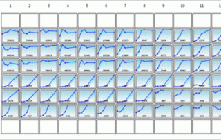
Generate growth curves over time and monitor cell counts and confluence at the individual well level.

Detection of Multiplexed GFP Reporters in Primary Articular Chondrocyte Cultures Using Cellometer Vision Image Cytometer

It's White Paper Wednesday! Read our featured white paper: Detection of Multiplexed GFP Reporters in Primary Articular Chondrocyte Cultures Using Cellometer Vision Image Cytometer In this work, we have developed an image cytometry method for detecting and monitoring the cell expansion and differentiation of articular chondrocytes in primary culture. First, the feasibility of utilizing image cytometry for detection of fluorescent is shown by comparing measured fluorescent positive cell populations to flow cytometry. Next, articular chondrocyte cultures were established in multi-well plates from either single or Cyan/eGFP double reporter mouse lines and grown for 20 days to test the utility of the [...]

By |2021-06-15T20:34:28+00:00February 25th, 2015|Categories: Cellometer, Cellometer Application News, Instrument|Tags: , , , , |0 Comments

Cellometer Vision uncovers new mechanisms by which Type I interferons direct autophagy in vitro

Introduction A diverse family of cytokines, Type I interferons (IFNs), is a group of proteins responsible for antiviral, antiproliferative, and immunomodulatory functions [1, 2]. Treating cells with Type I IFNs induces autophagy, a cellular recycling process that is considered a key cell survival strategy [3]. It is well known that Type I IFNs work through the JAK-STAT pathway, but recent evidence suggests that the MAP kinase pathway can affect the expression of IFN-regulated genes [4, 5]. Researchers demonstrated that IFN-induced autophagy affects cell cycle as well as cellular proliferation in a variety of cell lines. Because autophagy plays a major [...]

Cellometer Vision CBA image cytometry reveals the utility of the immunocompetent mouse model “Glowing Head” in assessing preclinical anti-tumor therapies

Introduction Preclinical models to evaluate therapeutic efficacy in the area of oncology have proven to be a challenge. The standard of preclinical assessment as always been immunosuppressed mice, but that model has not reliably predicted clinical efficacy [1]. Immunocompetent genetically engineered mice with tumor allograft models seem more promising, and yet reporter tags such as GFP and luciferase are targeted by competent immune systems, which may interfere with tumor growth and the response to therapeutics. Here, a collaborative group of researchers has created a reporter-tolerized genetically engineered mouse model, dubbed “Glowing Head” (GH), by targeting a luciferase-GFP reporter into the [...]

Iron oxide-based nanoparticles conjugated NuBCP-9 increase MCF-7 apoptosis as assessed by Cellometer Vision CBA Image Cytometer

Introduction The B-cell lymphoma-2 (BCL-2) family of proteins includes many key regulators of cell survival, cell death, and apoptosis. The deregulation of certain BCL-2 proteins is one of the first steps towards tumorigenesis and subsequent therapeutic resistance [1-4]. Targeting these BCL-2 family members is therefore an important area of oncology research [2, 4, 5]. Here, a collaboration of researchers has explored the targeting of these BCL-2 proteins with a BCL-2 binding peptide known as NuBCP-9 (FSRSLHSLL), which induces a conformational change in BCL-2 that alters the protein’s function from cell protection to cell killing. Researchers paired NuBCP-9 with iron oxide-based [...]

By |2021-06-15T20:35:46+00:00January 8th, 2015|Categories: Cellometer, Cellometer User Publications, Instrument|Tags: , |0 Comments

Investigation of IAPP Role in Increasing ROS Production and Apoptosis in p53-deficient Tumor Cells using Celigo Imaging Cytometer

The entire family of tumor protein p53 (TP53) enhances functions such as apoptosis and autophagy in normal cellular functioning. TP53 is a tumor repressor gene that is often inactivated in human cancers. Reactivating p53 has proven difficult to achieve therapeutically, however. Researchers at MD Anderson Cancer Center are investigating other members of the p53 pathway in order to elucidate new therapeutic options to suppress p53-deficient tumor growth. ΔN isoforms of two members of the p53 family, p63 and p73, are usually overexpressed in cancers and these isoforms (which lack the acidic transactivation domain) act on p53 in a dominant-negative fashion, [...]

Evaluation of Nanoparticle-Based Therapy for Triple-Negative Breast Cancer using Cellometer Vision CBA

Introduction to Nanoparticle-Based Therapy for Triple-Negative Breast Cancer Tia Harmon and Dr. Ruben Gonzalez-Perez from the Morehouse School of Medicine and Emory University utilized the imaging and analysis capabilities of the Cellometer Vision to investigate leptin peptide receptor antagonist-conjugated nanoparticles in order to inhibit leptin signaling, a key pathway that promotes growth and survival of triple-negative breast cancer (TNBC) cells. It was hypothesized that these nanoparticles would first stop the expression of leptin’s downstream target Notch, and would thereby increase the effectiveness of standard chemotherapeutic compounds in limiting tumor cell survival [1-4]. Materials and Methods A human ER+ cell line (MCF-7) [...]

By |2021-06-15T20:36:25+00:00December 8th, 2014|Categories: Cellometer, Cellometer User Publications, Immuno-oncology, Instrument|0 Comments

Introducing Teaching Lab Program with Cellometer Mini and Cellometer Auto 1000

In collaboration with our academic customers, we have developed a Teaching Lab program now available to any academic customer. Our program is designed to seamlessly integrate into courses that introduce students to: Using industry standard biotechnology tools and techniques Examine fundamental cell biology: cell health, morphology, viability Basic laboratory techniques: Hemacytometer vs. automated cell counting Hands-on experience using research tools in student-driven projects Incorporate novel technology into routine workflow Many colleges and universities nationwide have already incorporated our teaching lab program into their Freshman Research Initiatives and Research Methodology courses. The teaching lab program is a great teaching tool and [...]

Rapid Image Cytometry Method for Cell Counting and Viability Measurement for Cellular Therapy

Patients with cancers such as multiple myeloma, leukemia, lymphoma, and other metastatic cancers have tumor cells with unique immunological targets that when exploited, can lead to the complete destruction of the mass and a lasting remission from disease. Clinicians attack those targets using cell therapy, also known as “targeted immunotherapy”. Cell therapy is the programming of a patient’s own immune cells to target that patient’s tumor cells for destruction. One application of cell therapy, Adoptive Cell Transfer (ACT), employs select methods of genetic manipulation and propagation of those immune cells ex vivo so that the newly programmed cells can be [...]

Go to Top