Time-Course Monitoring of Primary Murine B1 and B2 Cell Proliferation using Cellometer Vision Image Cytometer

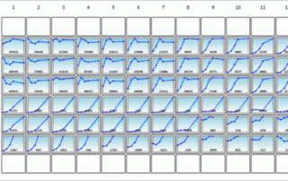
It's White Paper Wednesday! Read our featured white paper: Time-Course Monitoring of Primary Murine B1 and B2 Cell Proliferation using Cellometer Vision Image Cytometer Cell proliferation is an important assay for pharmaceutical and biomedical research to test the effects of a variety of treatments on cultured primary cells or cell lines [1, 2]. Previously, we have reported a rapid and accurate fluorescence-based cell population analysis method using a novel image-based cytometry system. The method is highly comparable to traditional flow cytometry using fewer cells [3-6]. Here we report the development of a novel method for the kinetic measurement of cell [...]

By |2021-06-15T19:50:01+00:00July 22nd, 2015|Categories: Cellometer, Cellometer Application News, Instrument|Tags: , , , , |0 Comments

Craft brewers enhance brewing process and product taste using the Cellometer X1 and X2 instruments

Avery Brewing Company in Boulder, Colorado has introduced a Cellometer to their brewing process, improving their procedures, as well as the product’s taste and quality. The Cellometer is employed as a quality control check over the yeast cells inside the facility’s fermenters. Making sure the yeast cells fermenting the beer are healthy and enzymatically active ensures a high quality product, with fewer mutated and/or dying cells that can negatively affect the beer’s flavor. Adam, the owner  of Avery Brewing Co. says that the Cellometer has “changed the way we made beer and it also changed the way our beer tastes”.   Watch this episode of [...]

Measuring Apoptosis and Necrosis Cell Populations by Heat and Compound Induction using the Cellometer Vision

It's White Paper Wednesday! Read our featured white paper: Measuring Apoptosis and Necrosis Cell Populations by Heat and Compound Induction using the Cellometer Vision Recently, a new imaging cytometry system has been developed by Nexcelom Bioscience LLC (Lawrence, MA, USA) for fluorescence-based cell population analysis [1, 2]. Originally, the system has been used for automated cell concentration and viability measurement using disposable counting slides with both bright-field (BR) and fluorescent (FL) imaging methods [3]. The system has recently been demonstrated to perform fast and accurate cell population analysis comparable to the traditional flow cytometry method. Download our white paper »

By |2021-06-15T19:50:43+00:00June 24th, 2015|Categories: Cellometer, Cellometer Application News, Instrument|Tags: , , |0 Comments

Comparison of Trypan Blue and Fluorescence-Based Viability Detection Methods Via Morphological Observation

It's White Paper Wednesday! Read our featured white paper: Comparison of Trypan Blue and Fluorescence-Based Viability Detection Methods Via Morphological Observation Determining cell viability is a vital component in many biological experiments that range from standard cell culture to the investigation of pharmacological agents on tumor cells. One of the earliest and most common methods for measuring cell viability is the trypan blue (TB) exclusion assay [1, 2]. Over the last two decades, there have been various publications on comparing TB exclusion and fluorescence-based cellular viability assays [1, 3-5]. Previous results have shown that in a time-course measurement, TB exclusion [...]

Measuring Physiological and Metabolic Characteristics of Yeast Health for Beer Fermentation Samples using the Cellometer Vision

It's White Paper Wednesday! Read our featured white paper: Measuring Physiological and Metabolic Characteristics of Yeast Health for Beer Fermentation Samples using the Cellometer Vision Characteristics such as the viability, vitality, glycogen, neutral lipid, and trehalose content of yeast samples must be measured to better understand changes in physiological and metabolic status during fermentation [4-9]. Monitoring changes of these parameters during fermentation can improve current production processes, and a subset of these parameters (glycogen, neutral lipid, and trehalose content) has been shown to play an important role in predicting yeast viability both during and after fermentation [10, 11]. Download our [...]

By |2021-06-15T19:52:03+00:00April 8th, 2015|Categories: Cellometer, Cellometer Application News, Instrument|Tags: , , , , |0 Comments

Comparing Fluorescence-Based Viability Detection Method using the Cellometer Vision

It's White Paper Wednesday! Read our featured white paper: Comparing Fluorescence-Based Viability Detection Method using the Cellometer Vision In this work, Cellometer Vision was employed to demonstrate rapid fluorescence-based viability measurements and the comparison of various fluorescent staining methods. First, fluorescent nucleic acid stains that examine membrane integrity were tested and validated by comparing against the standard trypan blue exclusion method. Similarly, fluorescent enzymatic stains that examine metabolic activities were tested and validated against the standard trypan blue exclusion method. Third, nucleic acid and enzymatic stains were compared by measuring viabilities of Jurkat cells incubated at different temperatures of water [...]

Cellometer Vision tracks apoptosis in embryonic neural stem cells after propofol exposure

Introduction Propofol, a widely used general anesthetic, has been shown to produce neurotoxicity in neonatal animal experiments [1-5]. The possible detrimental effects of anesthetics, particularly in developing brains, require further study, as recent studies suggest that general anesthesia given to children younger than four years of age can increase the chances of learning disabilities later in life [6]. Here, researchers used embryonic neural stem cells to investigate the neurotoxic effects of propofol in vitro. Materials and Methods Cell culture and drug exposure Embryonic neural stem cells were collected from embryonic day 16 Sprague-Dawley rats. Resulting cells were cultured in 96-well [...]

Bright field and Fluorescent Image Analysis for Screening Applications using Celigo

Generate growth curves over time and monitor cell counts and confluence at the individual well level.

Detection of Multiplexed GFP Reporters in Primary Articular Chondrocyte Cultures Using Cellometer Vision Image Cytometer

It's White Paper Wednesday! Read our featured white paper: Detection of Multiplexed GFP Reporters in Primary Articular Chondrocyte Cultures Using Cellometer Vision Image Cytometer In this work, we have developed an image cytometry method for detecting and monitoring the cell expansion and differentiation of articular chondrocytes in primary culture. First, the feasibility of utilizing image cytometry for detection of fluorescent is shown by comparing measured fluorescent positive cell populations to flow cytometry. Next, articular chondrocyte cultures were established in multi-well plates from either single or Cyan/eGFP double reporter mouse lines and grown for 20 days to test the utility of the [...]

By |2021-06-15T20:34:28+00:00February 25th, 2015|Categories: Cellometer, Cellometer Application News, Instrument|Tags: , , , , |0 Comments

Cellometer Vision uncovers new mechanisms by which Type I interferons direct autophagy in vitro

Introduction A diverse family of cytokines, Type I interferons (IFNs), is a group of proteins responsible for antiviral, antiproliferative, and immunomodulatory functions [1, 2]. Treating cells with Type I IFNs induces autophagy, a cellular recycling process that is considered a key cell survival strategy [3]. It is well known that Type I IFNs work through the JAK-STAT pathway, but recent evidence suggests that the MAP kinase pathway can affect the expression of IFN-regulated genes [4, 5]. Researchers demonstrated that IFN-induced autophagy affects cell cycle as well as cellular proliferation in a variety of cell lines. Because autophagy plays a major [...]

Go to Top