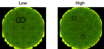
Ignyta tests Celigo against Cell Titer-Glo for cell proliferation

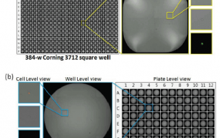
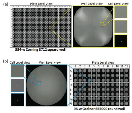
Here's a great example of how the Celigo image cytometer is able to perform common experiments while saving time and money! Ignyta, Inc. was looking for a new way to perform reagent-free proliferation analyses with suspension cells. This new method had to produce results which correlated well to their current method, Cell Titer-Glo®. Nexcelom and Ignyta partnered to perform a head-to-head cell proliferation comparison between Celigo® and Cell Titer-Glo. Using four suspension cell types (Ba/F3 parental cell line, Ba/F3 expressing an oncogenic gene, oncogenic gene mutant A and B), Ignyta plated all cells at a concentration of 5,000 cells/well in [...]

By |2021-06-15T19:47:39+00:00October 23rd, 2015|Categories: Celigo, Celigo User Publications, Instrument|Tags: , , |0 Comments

Live Webinar! Join us for “3D Tumor Spheroids Based Functional Assays”

Recent publications have suggested that using 3D tumor spheroids is a more predictive model for preclinical research. Nexcelom Bioscience has developed a standardized microplate method for rapid generation, imaging and analysis of 3D tumor spheroids using the Celigo image cytometer. 40 cancer cell lines' ability to form spheroids, optimal seeding densities and culture conditions have been established. Protocols measuring tumor growth, viability, migration and invasion have been utilized by many researchers for routine preclinical drug studies. If you are interested to learn more about how your research might benefit from working with 3D tumor spheroids, this is a great place to start! [...]

By |2021-06-15T19:48:33+00:00October 13th, 2015|Categories: 3D Models, Celigo, Celigo Application News, Instrument|Tags: , , |0 Comments

Increasing Efficiency in Cell Line Development using the Celigo Imaging Cytometer

Celigo Imaging Cytometer demonstrated the utility of automation in the development and monitoring of new CHO-based cell lines for increasing efficiency in cell line development.

A Rapid and Label-Free In Situ Assay Method for Cell Proliferation and Drug Toxicity using Celigo Imaging Cytometer

It's White Paper Wednesday! Read our featured white paper: A Rapid and Label-Free In Situ Assay Method for Cell Proliferation and Drug Toxicity using Celigo Imaging Cytometer In this study, Celigo was used to screen a compound library for effects on cell proliferation in adherent and non-adherent cell lines. Human lung carcinoma (A549) and promyelocytic leukemia (HL-60) cells were treated with a panel of compounds to inhibit proliferation. Finally, the Celigo system used image-based analysis to measure changes in cell morphology upon compound treatment. These data indicate that certain anti-proliferative compounds can have secondary effects on cell health or physiology, [...]

3D Tumor Spheroid Analysis Method for HTS Drug Discovery using Celigo Imaging Cytometer

It's White Paper Wednesday! Read our featured white paper: 3D Tumor Spheroid Analysis Method for HTS Drug Discovery using Celigo Imaging Cytometer U87MG cells were used to create tumorspheres in 384-well plates that were subsequently analyzed by imaging. The data illustrate that reproducible 3D spheroids can be formed in 384-well plates. Fluorescent viability studies were carried out with the imager using pixel intensity analysis. Moreover, the assay was validated for drug screen using various drug compounds that have shown anti-proliferative effects. Together, these data demonstrate that the tumorsphere formation assay can be developed, validated and used for high-throughput anti-cancer compound [...]

Non-Disruptive Quantification of 2° Reprogrammed iPS Colonies Using Celigo Imaging Cytometer

Celigo  imaging cytometer has been applied to provide automated, rapid assessment of iPSC reprogramming.

Automated Method for Determination of Infectious Dose (TCID50) using Celigo Imaging Cytometer

It's White Paper Wednesday! Read our featured white paper: Automated Method for Determination of Infectious Dose (TCID50) using Celigo Imaging Cytometer Nexcelom's Celigo imaging cytometer has been applied to provide automated, rapid assessment of viral infectivity in a range of plate formats [4]. Using f-theta optics, Celigo provides high quality, whole-well images using bright field and/or fluorescent illumination. Automated segmentation and analysis provide quantitative and objective output of CPE based on characteristic changes to the host cell monolayer. Download our white paper »

Characterization of Breast Cancer Drugs via Mammosphere Morphometric Analysis Using Celigo Imaging Cytometer

It's White Paper Wednesday! Read our featured white paper: Characterization of Breast Cancer Drugs via Mammosphere Morphometric Analysis Using Celigo Imaging Cytometer A panel of cytotoxic drugs, including doxorubicin and paclitaxel were used to study their effects on various breast cancer cell lines such as MDA-MD-436, MCF-7, SKBR3 and MDA-MB-231. Results show that the Colony Counting application can also be used to evaluate the clonogenicity and self-renewal of cancer stem/tumor-initiating cells by automatically analyzing mammosphere populations. The Colony Counting application on Celigo provides an efficient, reproducible and automated method for assessing the number, size, and morphology of cancer spheroids within [...]

Nexcelom’s Celigo Detects CRISPR Transfection Efficiency of sgRNA in CHO Cells

Novel web-based tool ensures efficient, fast, and low-cost genetic manipulation of CHO cells for future biopharmaceutical applications.

Investigation of IAPP Role in Increasing ROS Production and Apoptosis in p53-deficient Tumor Cells using Celigo Imaging Cytometer

The entire family of tumor protein p53 (TP53) enhances functions such as apoptosis and autophagy in normal cellular functioning. TP53 is a tumor repressor gene that is often inactivated in human cancers. Reactivating p53 has proven difficult to achieve therapeutically, however. Researchers at MD Anderson Cancer Center are investigating other members of the p53 pathway in order to elucidate new therapeutic options to suppress p53-deficient tumor growth. ΔN isoforms of two members of the p53 family, p63 and p73, are usually overexpressed in cancers and these isoforms (which lack the acidic transactivation domain) act on p53 in a dominant-negative fashion, [...]

Go to Top