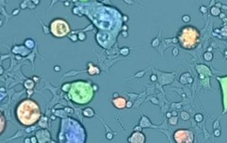
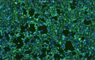
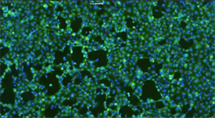
What is Image Cytometry?
Image cytometry and how it compares to other methods. Improve the quality of cell counting results to increase confidence in processes and workflows.
Image cytometry and how it compares to other methods. Improve the quality of cell counting results to increase confidence in processes and workflows.
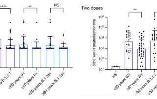
With new variants and waning immunity occurring, especially in elderly and immunocompromised individuals, it is important to understand how to utilize the tools we have to prevent severe disease and death.
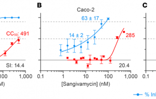
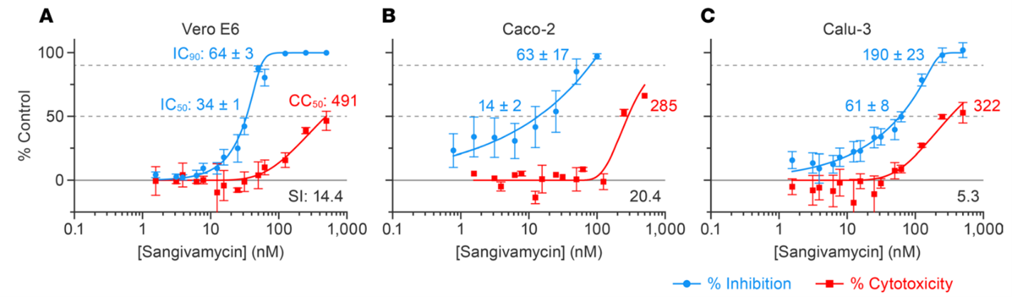
Sangivamycin is an unsuccessful anti-cancer drug candidate that has proven to be a potent inhibitor of multiple viruses. The authors of this study hypothesized that this compound would also be active against SARS-CoV-2.
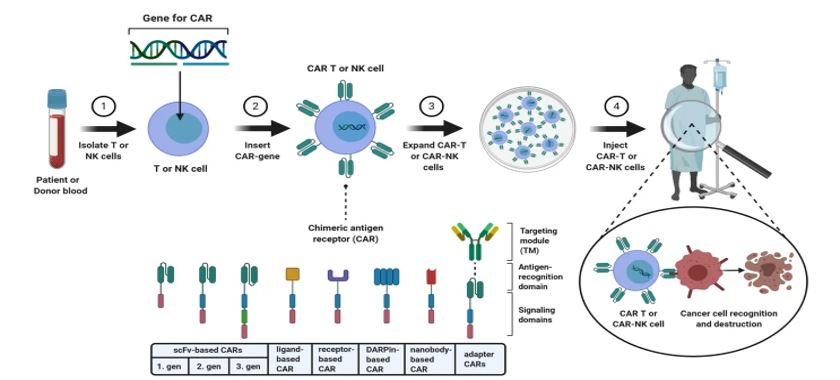
For the development of new drugs and the advancement of new cell therapies, pharmaceutical companies have recurred to cytotoxicity assays as a quick way to assess the viability of cell lines in response to an external stimulus.
Acute respiratory distress syndrome (ARDS) is characterized by pulmonary edema, often causing lung failure or death.
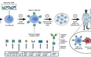
Drug repurposing, also known as drug repositioning is a powerful tool for the rapid expansion of available therapy options by identifying new therapeutic uses from pre-existing drugs.
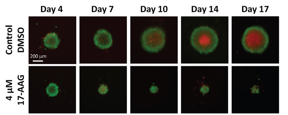
Image-based, automated analysis of 3-D tumor spheroids in microwell plates is becoming standard practice for the evaluation of anticancer drug compounds.
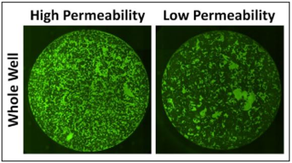
It's White Paper Wednesday! Read our featured white paper: Automated Method for Determination of Infectious Dose (TCID50) using Celigo Imaging Cytometer Nexcelom's Celigo imaging cytometer has been applied to provide automated, rapid assessment of viral infectivity in a range of plate formats [4]. Using f-theta optics, Celigo provides high quality, whole-well images using bright field and/or fluorescent illumination. Automated segmentation and analysis provide quantitative and objective output of CPE based on characteristic changes to the host cell monolayer. Download our white paper »

