Accurate Cell Counting is Important for ELISpot
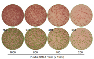
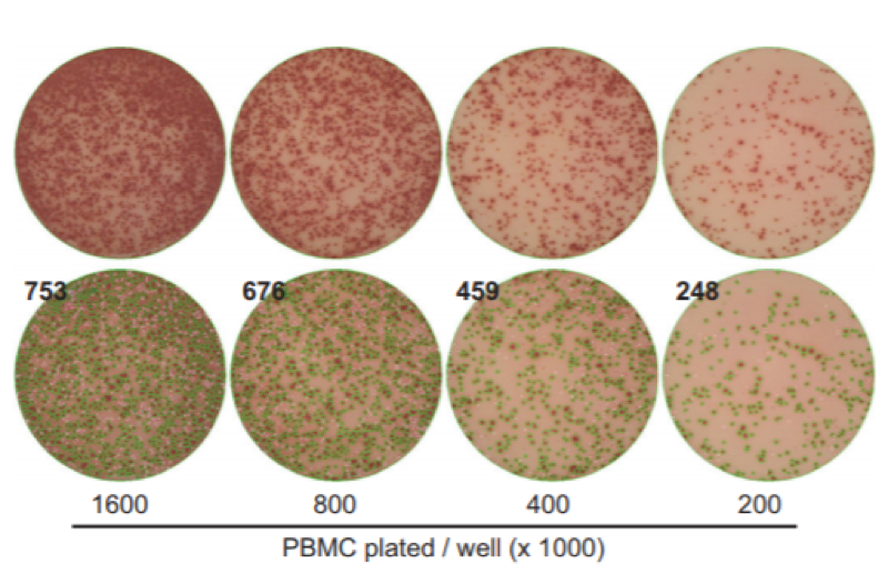
ELISpot is an assay for the detection and enumeration of cytokine secreting cells. Accurate cell counting is important for ELISpot.
ELISpot is an assay for the detection and enumeration of cytokine secreting cells. Accurate cell counting is important for ELISpot.
If you rely on a flow cytometer in a core lab or a central facilities lab, have you ever wondered if there was a better way? Have you ever wished to have full control over your experiments, rather than hand your sample over to a technician? There was an interesting article written recently about the needs of researchers. Have you seen this? Those interviewed for the article suggest that there are some main factors for consideration: The instrument size needs a smaller footprint than existing products offer The price of the instrument should be under $100,000 Software ease-of-use to reduce [...]
What is GFP? Green Fluorescent Protein (GFP) is a 26.9 kDa protein first identified in crystal jellyfish, Aequorea victoria. It was discovered that when exposed to blue or ultraviolet light the protein fluoresces green. After GFP was first expressed in E. coli in 1994 it was soon confirmed that GFP can also be successfully expressed in other organisms as well. Since then, not only have many fluorescent proteins of different colors been generated, but their function is enhanced to provide a faster and stronger fluorescent signal. GFP Applications GFP is often used as a reporter of gene or protein expression. [...]
Jurkat cells were used to analyze cell cycle kinetics following treatment with the cell-cycle-arresting drug etoposide. Etoposide is designed to arrest the cells at the G2 phase of the cell cycle. Jurkat cells were incubated with media only (control) or etoposide (0.06 µM, 0.12 µM) for 24 hours. Control and drug-treated cells were ethanol fixed and stained with cell cycle propidium iodide reagent. For each sample, 20 µl of cell sample (at ~4 x 10⁶ cells / mL) was loaded into a Cellometer imaging chamber, inserted into the Vision CBA Analysis System, and imaged in both bright field and fluorescence. [...]
Introduction One of the most common and popular methods for cell cycle detection is the use of fluorescence-based dyes. There are a number of fluorescent-based dyes that are capable of binding to double stranded DNA upon cell fixation. Propidium iodide (PI) and DAPI are two such dyes. Since the amount of bound fluorescent dye is directly proportional to the amount of DNA present within a cell, these dyes can be used to detect the cell cycle within a population of cells. How it works: As the cells are going through the cell cycle, the amount of DNA that is contained [...]
Calcein AM (Calcein acetoxymethyl ester) is a non-fluorescent compound that passively enters cells. In metabolically active cells, Calcein AM is converted by cytosolic esterases into green fluorescent Calcein. The fluorescent Calcein is retained by live cells with intact membranes. Only cells possessing active cytosolic esterases fluoresce green. This allows for quick and easy detection of metabolically-active (vital) cells in a sample. […]
GFP and Determination of Transfection Efficiency Green Fluorescent Protein (GFP) is a 26.9 kDa protein that fluoresces bright green when exposed to blue or ultraviolet light. GFP was first identified as a protein and extracted from the Aequorea victoria jellyfish in 1962 by Osamu Shimomura, et al. The GFP protein was first cloned in 1992 and it was soon confirmed that GFP protein expressed in other organisms generates fluorescence. An area within a cell or tissue is briefly illuminated, causing the GFP protein to fluoresce, allowing GFP-tagged proteins to be identified. In 2008, Osamu Shimomura, Marty Chalfie, and [...]
Autophagy is a normal process by which eukaryotic cells break down out-dated and damaged cellular organelles and proteins to be replaced with new ones. It is also a survival mechanism providing cells with energy and substrates for cellular processes in times of stress and starvation. Autophagy is a multi-step process involving initiation, formation of autophagosomes (vesicles that capture and deliver cytoplasmic material to lysosomes for digestion), maturation, and degradation (Figure 1.) […]
Caspase-3 belongs to a family of evolutionally conserved cysteine proteases that play a key role in regulating programmed cell death, or apoptosis, a normal process required for maintenance of tissue homeostasis and the regulation of physiological functions1. The two main apoptosis activation pathways are the extrinsic and the intrinsic pathways. The extrinsic pathway is activated by the binding of ligands (including TNFα, FasL, and TRAIL) to cell-surface receptors. The intrinsic, or mitochondrial, pathway is typically activated in response to DNA or cellular damage. The convergence of the extrinsic and intrinsic pathways occurs at the proteolytic activation of caspase-34. Once caspase-3 is [...]
Caspase-8 Caspase-8 belongs to a family of evolutionally conserved cysteine proteases that play a key role in regulating programmed cell death or apoptosis. The two main apoptosis activation pathways are the extrinsic and the intrinsic pathways. The extrinsic pathway is activated by the binding of ligands (including TNFα, FasL, and TRAIL) to cell-surface receptors and the conversion of procaspase-8 to the active caspase-8 protease.1 Caspase-8 quickly converts procaspase-3 to active Caspase-3, facilitating many of the cellular and biochemical events of apoptosis. The intrinsic, or mitochondrial, pathway is typically activated in response to DNA or cellular damage and begins with the [...]