What is Image Cytometry?
Image cytometry and how it compares to other methods. Improve the quality of cell counting results to increase confidence in processes and workflows.
Image cytometry and how it compares to other methods. Improve the quality of cell counting results to increase confidence in processes and workflows.
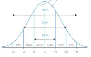
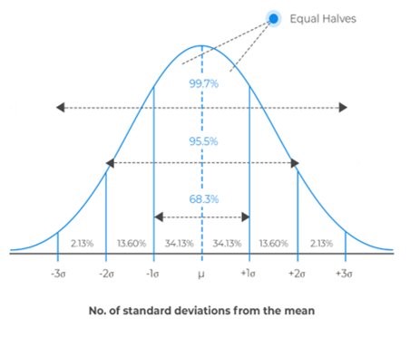
“Biological variation” is an easy explanation to reach for when repeated measurements don’t match up very well. It is vague enough to excuse inconsistency in nearly any biological experiment, and in many cases, it is difficult to disprove.
Download the Poster Development of an image-based HCS compatible method for endothelial barrier function assessment Oleksii Dubrovskyi, Erica Hasten, Steven M. Dudek, Michael T. Flavin, and Leo Li-Ying Chan Acute respiratory distress syndrome (ARDS) is a serious condition with high mortality rate that has increased in the recent years due to the COVID-19 pandemic. Currently, effective pharmacological therapies have yet been discovered for ARDS, despite decades of laboratory and clinical studies. ARDS onset typically generates an increase in the endothelial permeability causing the development of pulmonary edema that leads to respiratory failure during the primary event. In this work, we [...]
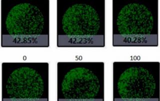
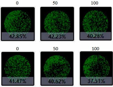
Perform simultaneous imaging and analysis to rapidly capture and quantify spheroid images in every well with advanced software segmentation.
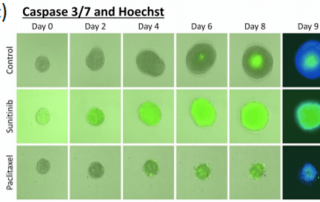
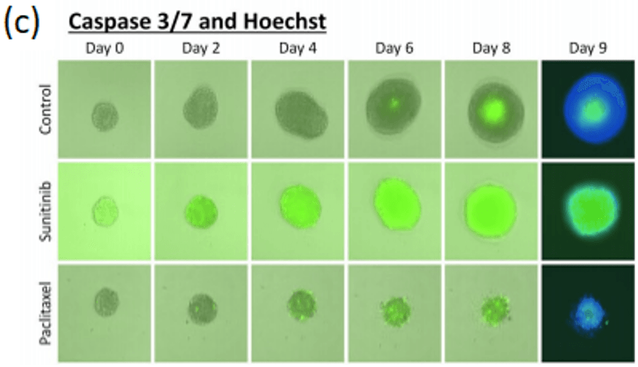
Download the Poster Apoptosis Analysis of Jurkat Cells using the Cellometer Vision Leo L. Chan, Tim Smith, Alnoor Pirani, Emily Lyettefi, Ning Lai, Jean Qiu, and Bo Lin We demonstrate a rapid and cost-effective method for apoptosis analysis at various stages of Jurkat cells using the Cellometer® Vision. This method has the ability to eliminate many known issues caused by manual hemacytometer and flow cytometer. By using Cellometer® Vision, the assay time for obtaining apoptosis result is greatly reduced, which is significant for research development in academia and industry. The current methods for apoptosis analysis utilizes standard flow cytometry, but [...]
Download the Poster A Novel Imaging Cytometry Method for Immunophenotyping Leo L. Chan, Xuemei Zhong, Jean Qiu, Peter Y. Li, Bo Lin The ability to detect and enumerate cells with specific surface markers is essential for clinical diagnosis and biomedical research. The traditional cell analysis instrumentation for immunophenotyping involves the use of fluorescence microscopy, confocal fluorescence scanning, and flow cytometry. However, these systems are often expensive to purchaseand maintain. In this work, we demonstrate a novel low-cost device, Cellometer® system, which combines both microscopic imaging and cytometry methods with comparable detection sensitivity. This method utilizes both bright-field and fluorescence imaging [...]
Download the Poster A Rapid Alternative Method for Cell Cycle Analysis Using Cellometer Vision Leo L. Chan, Xuemei Zhong, Jean Qiu, Peter Y. Li, and Bo Lin Traditional cell cycle analysis instrumentation involves the use of fluorescence microscopy, laser scanning cytometry, and flow cytometry. In this work, we demonstrate new applications for the Cellometer Vision, which utilizes an imaging cytometry method for cell cycle analysis. This method employs both bright-field and fluorescence imaging of a disposable cell counting chamber to quickly provide concentration and percentages of cell subpopulations. Signal measurement and data analysis could be performed less than fivemin. Cell [...]
Effective long-term cryopreservation of human mesenchymal stem cells is an essential component of regenerative medicine and tissue engineering applications.
Many factors and considerations must be optimized to develop the appropriate cell-based assays for gene therapy development.
View these simple step-by-step instructions for counting cells using a hemoctyometer and microscope. Includes the formula for calculating the dilution factor.

