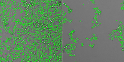
Cytopathic effects of SV-5 virus on CHO-K1 cells measured by morphological analysis
In this experiment, the goal is to measure the time-course CPE of SV-5 virus on the CHO-K1 host cells in order to determine the earliest time point that CPE can be automatically detected.
The CHO-K1 host cells are seeded in 6-well microplates, incubated and allowed to adhere overnight. The host cells are inoculated with SV-5 virus as the positive sample and media for the negative control. The 6-well microplates are scanned at 10% of the well that required less than 1 min/plate, and analyzed using Celigo from day 0 to 12. The CHO-K1 cell morphology are analyzed directly in bright field imaging to determine the CPE.

Time-course bright field and analyzed images (aspect ratio: round vs. elongated cells) for the positive SV-5 virus. The decrease in elongated cells % of CHO-K1 cells indicates cytopathic effects.
Time-course bright field and analyzed images (aspect ratio: round vs. elongated cells) for the negative media control.

Time-course aspect ratio percentages results, showing the CPE occurring on day 5.
Example CPE measurement performed using the Celigo Image Cytometer:
CPE of SV-5 virus on CHO-K1 cells
Example CPE measurement performed using the Celigo Image Cytometer:
CPE of SV-5 virus on CHO-K1 cells
The Celigo Image Cytometry system performs high-througput, whole-well imaging and quantitative data in bright field and up to four fluorescent channels for a wide variety of cell-based assays.
Learn more about modern virology assays: