Automated focus reduction neutralization test using GFP-expressing respiratory syncytial virus (RSV) with hamster serum
- The host cells are seeded in a 24-well microplate and incubated for 100% confluence
- The cells are then infected by GFP-RSV and incubated with low, medium, and high hamster sera concentrations
- Finally, the plate is scanned and analyzed with Celigo to count the number of foci in each well

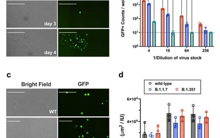
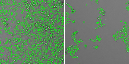
Zoomed in bright and fluorescent images of GFP-RSV infected foci

Plate-view of the 24-wells showing reduction in number of GFP foci at high concentration of serum
The Celigo Image Cytometer automates antibody neutralization assays:
Automated focus reduction neutralization test using GFP-expressing RSV
The Celigo Image Cytometry system performs high-througput, whole-well imaging and quantitative data in bright field and up to four fluorescent channels for a wide variety of cell-based assays.
Learn more about modern virology assays: